
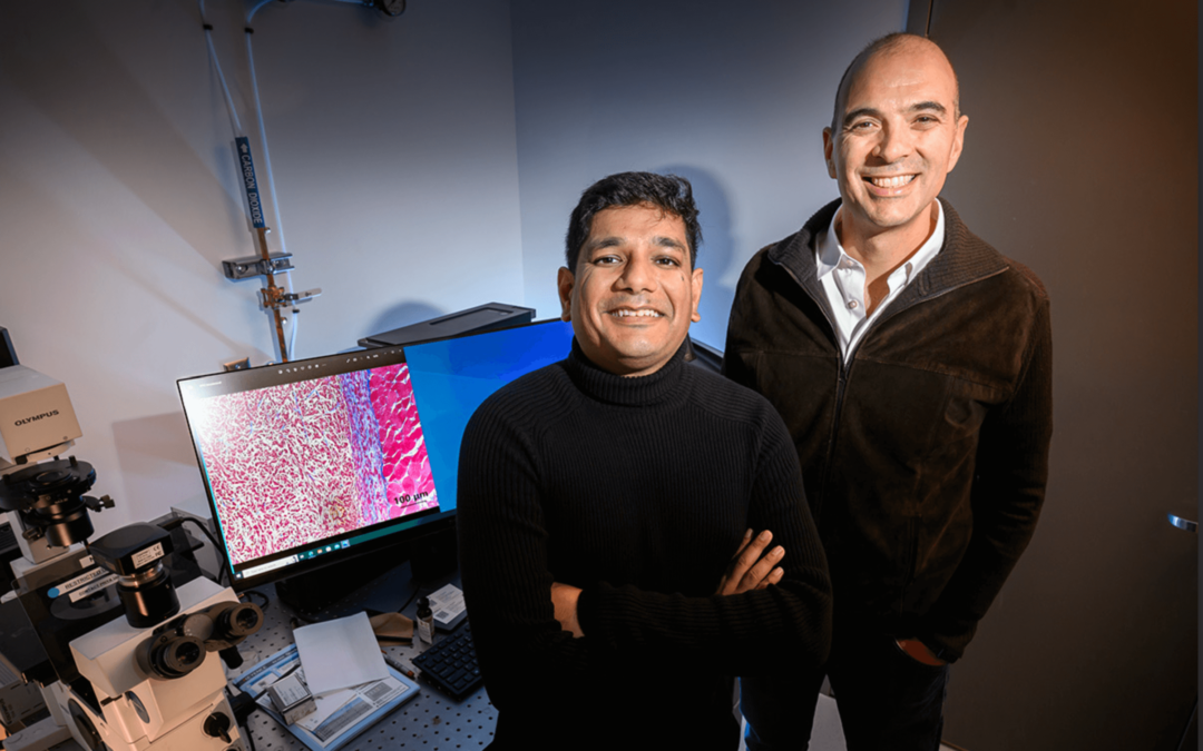
Postdoctoral researcher Indrajit Srivastava, left, and CCIL researcher Viktor Gruev, professor of electrical and computer engineering, led a team of researchers developing new cancer imaging agents that can light up two cancer biomarkers when lit by one fluorescent wavelength.
Cancer surgeons may soon have a more complete view of tumors during surgery thanks to new imaging agents that can illuminate multiple biomarkers at once, developed by Cancer Center at Illinois (CCIL) researchers. The fluorescent nanoparticles, wrapped in the membranes of red blood cells, target tumors better than current clinically approved dyes and can emit two distinct signals in response to just one beam of surgical light, a feature that could help doctors distinguish tumor borders and identify metastatic cancers.
The imaging agents can be combined with bioinspired cameras, which the researchers previously developed for real-time diagnosis during surgery, said research group leader Viktor Gruev, a CCIL member and professor of electrical and computer engineering. In a new study in the journal ACS Nano, the researchers demonstrated their new dual-signal nanoparticles in tumor phantoms – 3D models that mimic the features of tumors and their surroundings – and in live mice.
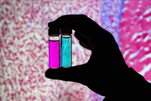
Researcher Indrajit Srivastava holds solutions of nanoparticles that can target two cancer biomarkers, giving off two distinct signals when lit by one fluorescent wavelength. This could give surgeons a more complete picture of a tumor and guide operating-room decisions. In the background is a microscopic scan of a tissue sample. (Photo by Fred Zwicky)
“If you want to find all the cancer, imaging one biomarker is not enough. It could miss some tumors. If you introduce a second or a third biomarker, the likelihood of removing all cancer cells increases, and the likelihood of a better outcome for the patients increases.” said Gruev, who also is a professor in the Carle Illinois College of Medicine. “Multiple-targeted drugs and imaging agents are a recent trend, and our group is driving the trend hard because we have the camera technology that can image multiple signals at once.”
Traditionally, a surgeon removes a tumor and sends it to a pathologist for assessment, a process that can take hours to days, said Illinois postdoctoral researcher Indrajit Srivastava, the first author of the paper. As research has moved toward real-time diagnostics, several challenges have prevented wide application: Many tumor-targeted imaging agents only minimally reach their tumor targets, instead being quickly cleared from the blood stream and accumulating in the liver, Srivastava said.
“A few people before us have used nanoparticles coated with red blood cells and found they circulate longer – a few days. We saw the same thing in our mice: The membrane-coated nanoparticles circulated longer in blood, with reduced uptake in the liver. Because they were circulating longer, more of the imaging agents accumulated in the tumors, giving us a stronger fluorescent signal,” Srivastava said.
The two biomarkers targeted by the new imaging agents include one that is prevalent in early cancer and one that is prevalent in late-stage cancer, which is more likely to be metastatic. The researchers found that the probes were effective at distinguishing cancerous tissue from healthy tissue, as well as distinguishing the two signals from each other.
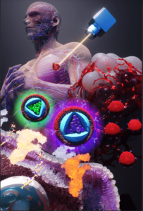
One wavelength of laser light during surgery elicits two distinct signals from the membrane-wrapped nanoparticles, one color for each of the two biomarkers that the nanoparticles target. One is common in earlier stages of cancer, and the other indicates later-stage – and possibly metastatic – cancer. (Image by Ella Maru Studios)
“This is appealing for surgical application, as it could help determine where exactly to make the cut. Having multiple signals gives a more overall picture of the tumor. And it could tell a surgeon, ‘This may be metastatic, you may want to be more aggressive in your removal.’” Srivastava said.
Only needing one wavelength of laser light to elicit multiple signals is another benefit for surgical applications, as it makes the instrumentation much more compact than those requiring multiple lasers for each needed wavelength, Gruev said.
The researchers plan to develop more tumor-imaging agents that target multiple markers, and to move forward with further preclinical and clinical studies using their dual-signal dyes with surgical goggles they have developed.
“In this battle for ensuring we remove all the cancer cells during surgery, we need investments both in the imaging camera technology and in the tumor targeting agents,” Gruev said. “This work is helping us better realize and guide the holistic approach that we are taking as we are getting closer and closer to clinical trials.”
The U.S. Congressionally Directed Medical Research Programs, the National Institutes of Health and the National Science Foundation supported this work. Gruev also is affiliated with the department of bioengineering and the Beckman Institute for Advanced Science and Technology.
Editor’s notes:
To reach Viktor Gruev, email vgruev@illinois.edu.
To reach Indrajit Srivastava, email indrajit@illinois.edu.
The paper “Cell-membrane coated nanoparticles for tumor delineation and qualitative estimation of cancer biomarkers at single wavelength excitation in murine and phantom models” is available online.
DOI: 10.1021/acsnano.3c00578
The original story featured on the Illinois News Bureau on June 5, 2023, written by Liz Ahlberg Touchstone,