
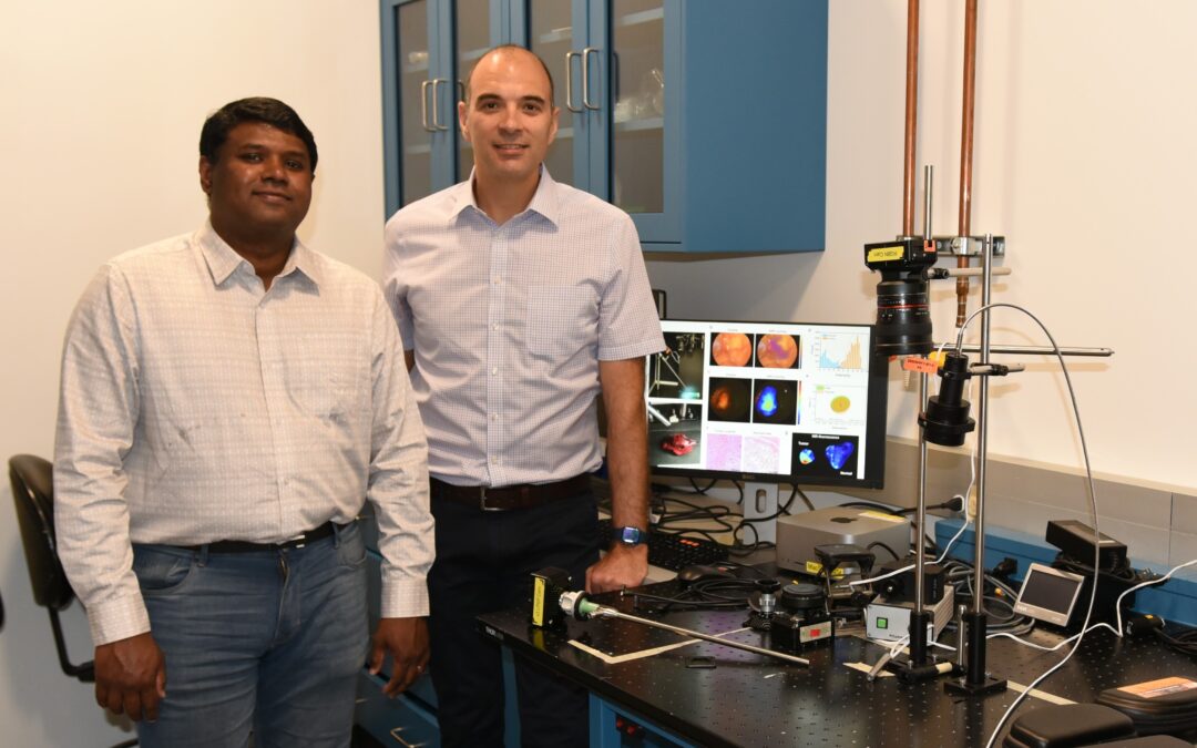
Graduate Student Mebin George and CCIL Member Viktor Gruev with their novel endoscopic imaging system.
Researchers from the Cancer Center at Illinois (CCIL) traveled the rewarding journey from benchtop to the operating room, observing their novel technology in the hands of surgeons treating lung cancer patients. Building upon their lab’s novel bioinspired imaging technology, the team led by the CCIL’s Viktor Gruev, professor of electrical and computer engineering, developed an improved endoscopic imaging system that achieves a 60% improvement upon existing FDA-approved endoscopic systems.
Endoscopy is emerging as a standard of care for cancer procedures; however, the standard ‘white light’ endoscopic technology falls short when it comes to seeing cancer cells. To remedy this problem, Gruev’s team designed a fluorescence-guided endoscopic imaging system that integrates their mantis shrimp-inspired hexachromatic image sensor, a novel rigid endoscope customized for near-infrared (NIR) color imaging, and a custom illumination fiber bundle. Fluorophores – dyes in this case – are a fluorescent chemical compound that tags the cells and then sends light back when excited by photons.
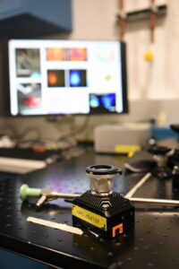
Bioinspired camera used with an endoscope for imaging multiple tumor targeted probes. The system was tested in the operating room on ex vivo samples from patients with lung cancer.
The bioinspired system goes beyond traditional endoscopic cancer imaging technology completing an impressive trifecta. Using multiple NIR fluorophores, the novel systems images multiple tumor-targeted probes, quantifies cancer cells, and detects tumor-specific biomarkers in ex vivo samples.
“We were not content to simply enhance the surgeon’s vision of the cancer cell landscape. We went a step further to identify biomarkers at the tumor environment to give clinicians more valuable information for stronger decision-making,” said Mebin Abu George, first author of the teams’ published research.
Because many cancers are heterogeneous rather than homogenous, the ability to see and interpret varied biomarkers gives the imaging system – and surgeons – a real advantage.
“The holy grail in cancer surgeries is to remove all cancer cells. We are moving into an age where there is a need to image multiple tumor-targeted drugs for this aim. Our lab is at the forefront of designing imaging technology for this goal; we are the only ones who can image multiple near-infrared biomarkers. The challenge remains with chemists seeking regulatory approvals so that the drugs can make it into the operating room where we are waiting to image them to eradicate all the cancer tissue,” said Gruev.
Gruev’s lab has shown success for over a decade in developing multispectral cameras. They have seen the fruits of their labor move into clinical trials; however, this is the team’s first venture into minimally invasive testing with endoscopy. With funding from the CCIL, the research team traveled weekly to the University of Pennsylvania to work with collaborators to implement the new imaging system in the clinic.
“The icing on the cake was our clinical study at the University of Pennsylvania, where we took the system into the operating room and saw the surgeons performing a cancer resection. The surgeon handed the specimen to us. It reminded us of what we are here for – we are not just developing some system in theory; we want to see clinical translation,” said George.
What comes next for the research team?
“Thanks to the support grant from the CCIL, as an extension of this project, we want to take our imaging technology beyond community hospitals and cancer centers in the U.S. to places like Benin, Africa, where we have a partnership. We want to innovate clever, cost-effective designs for those who have fewer resources,” said Gruev.
While the team reached a milestone with this new technology’s design, development, and implementation, they will continue to pursue additional clinical studies to address other types of cancer. The team’s research is published in the paper “Bioinspired Color-Near Infrared Endoscopic Imaging System for Molecular Guided Cancer Surgery” in Journal of Biomedical Optics (JBO).
—
Editor’s notes:
This work was performed in collaboration with Dr. Sunil Singhal at the University of Pennsylvania. It was funded by grants from the U.S. Air Force Office of Scientific Research (FA9550- 18-1-0278), Congressionally Directed Medical Research Programs (W81XWH-19-1-0299), National Science Foundation (2030421), and National Institute of Health (1P01CA254859), and supported by funding from the Cancer Center at Illinois (CCIL).
The paper is available here: doi.org/10.1117/1.JBO.28.5.056002
This story was written by Jonathan King, CCIL Communications.