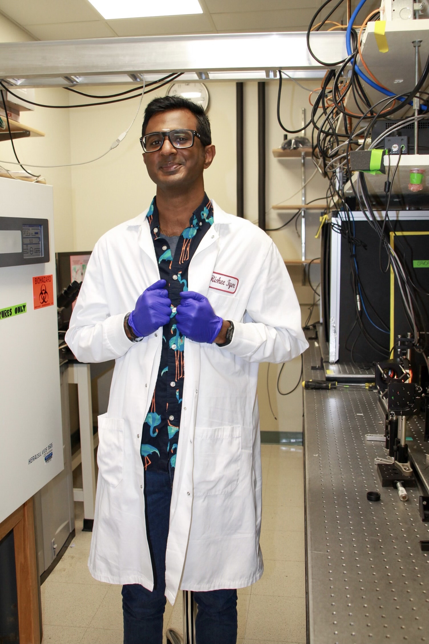
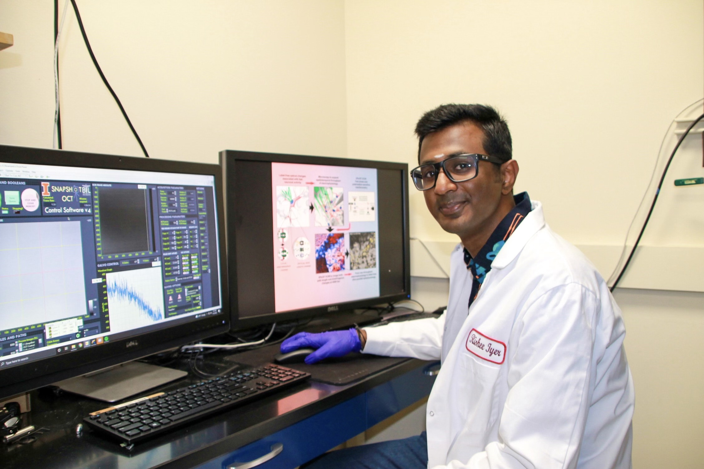
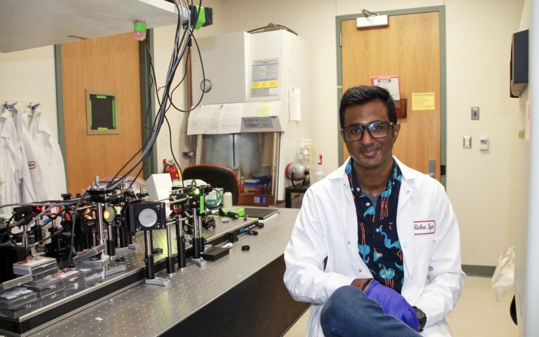
Rishee Iyer, Electrical and Computer Engineering Graduate Student
Tissue Microenvironment (TiMe) Program, 2022 Cohort
What does your educational path look like?
I did my undergraduate degree in India in electrical and electronics engineering, but I wasn’t too sure what I wanted to go into. I started doing this project where I met with a new professor, and he asked me to go into biometrics. I got into bioengineering when I accidentally stumbled upon a biomedical Wikipedia page.
Then, I did my masters in biomedical engineering at Cornell University. During my masters, I became more interested in the neuro microenvironment after taking a couple of neurophysiology classes.
What first sparked your interest in science?
I’ve always been super curious. As a child, I was always asking “why?” I wanted to be an astronaut but then I realized that I would never pass the physical training. I decided to stick to science, and I studied engineering; now I’m doing research.
Why is cancer research important to you?
Cancer is one of the leading causes of death, but we still know so little about it, especially in terms of diagnostics. All the research we work on in Dr. Stephen Boppart’s lab is important because it can accelerate diagnosis non-invasively.
How has being in the TiME program helped you grow?
One of the hard parts about doing research in an interdisciplinary environment is that you’re sort of stuck between these two worlds. Being able to find this community of students doing similar research is an invaluable thing, and that’s what the TiME program has provided me with.
What kind of research are you doing?
I’m working on imaging nerve cells using optical microscopy. If we think about how we would measure the electrical activity of nerve cells, we might imagine poking at them with an electrode, which is very invasive and not clinically translatable at all. The other way to measure the electrical activity is to add dyes and fluorescent molecules to it, which modifies the environment and therefore is also not clinically relevant.
We know that there are fundamental tools that we must develop to be able to see these neurons in action without adding any labels and non-invasively. I’m working on this research because optical microscopy is practically harmless, and its clinical relevance is starting to increase every day. I want to look at these neurons and their action without harming them.

What sort of impact are you hoping your research will have on society?
The tools that we are developing would enable us to go from the lab to the bedside. That is the impact that I want to make: I want to build these tools that are robust and versatile so that they can be applied in multiple places and make them easy to understand and use.
What are some challenges that you are currently facing in your research?
To be able to clinically translate something, it has to be a versatile technique that is accessible to all and harms as few people as possible, and two major things that we have to consider are safety and ethics.
What is it like to be under the mentorship of your supervisor?
My supervisor is Stephen Boppart, professor of electrical and computer engineering. The best part about working with him is that he lets his students do their research independently and he encourages us to come up with new ideas, but also to make executive decisions on how these research projects move forward.
What do you hope to do in the future?
I’ve always been interested in academia, and I love teaching. I do want to look into that, so I will consider a postdoctoral fellowship or a faculty position.
Outside of your research, what are some of your personal interests?
I do a lot of science-themed oil paintings. I also create a lot of science communication comic books and stuff like that. I’m very interested in graphic design, comic books, and painting.

– Written by Lisa Mei, Cancer Center at Illinois Communications Intern

