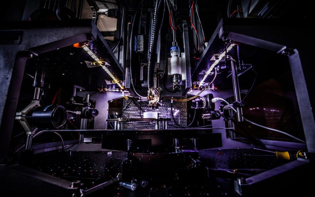
Urbana, Ill. – The Chemical Imaging and Structures Laboratory at the University of Illinois focuses on chemical imaging to improve our understanding of the pathology of many diseases, including cancer. Led by the Director of the Cancer Center at Illinois and founder professor in engineering, Rohit Bhargava, several graduate students and postdoctoral research fellows have dedicated their time to designing artificial intelligence (AI) methods to facilitate the development of infrared spectroscopy.
When imaging tissues with infrared spectroscopy, the chemical bonds that exist in biological components such as cancer cells vibrate at different frequencies, which can be captured as absorption patterns or signatures. Using this method, researchers can collect millions of signatures and separate the data to differentiate between healthy and diseased cells.
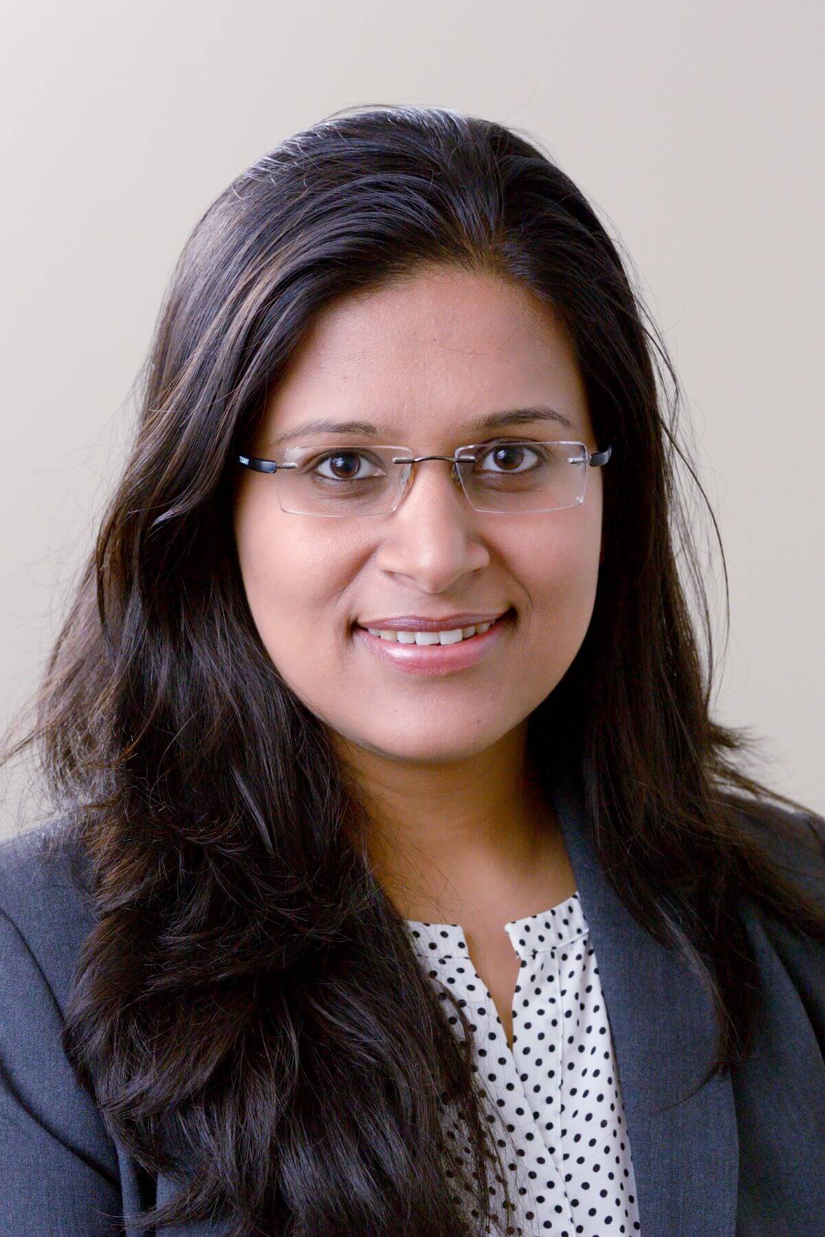
Shachi Mittal, former Illinois postdoctoral fellow and current assistant professor of chemical engineering at the University of Washington, specialized in obtaining high-definition IR data from breast cancer biopsies and developing traditional machine learning approaches like random forest (RF) algorithms and deep learning models.
“When a diagnosis is made, it’s usually made by a pathologist who consults multiple experts before making a decision. RF algorithms are like forests with many trees, where each tree mathematically predicts what the data represents,” said Mittal. “With 500 trees in the forest, we can get the ‘majority vote’ and reduce the uncertainty of the prediction.”

Shachi Mittal.

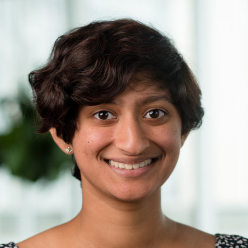
Serena Abraham.
New Ph.D. student, Serena Abraham, is continuing Mittal’s work in applying deep learning (DL) techniques to IR data to improve cancer diagnostics with a particular focus on breast cancer classification. By leveraging spatial and spectral information, Abraham looks to facilitate speedy diagnoses and super-resolution clinical imaging. Abraham is also assisting in a clinical collaboration with surgical oncologist, Anna Higham (M.D., Carle Foundation Hospital), on a project developing infrared spectroscopy for use in operating rooms. *
“I started my studies in genomics and image-based modeling, and I’m using the same AI tools here in the Bhargava lab. These tools are applicable to almost every field where analysis is required as a predictive tool to understand how systems behave — and at its core, the human body is an extremely complex stochastic nonlinear system that we try to approximate in research,” Abraham said.
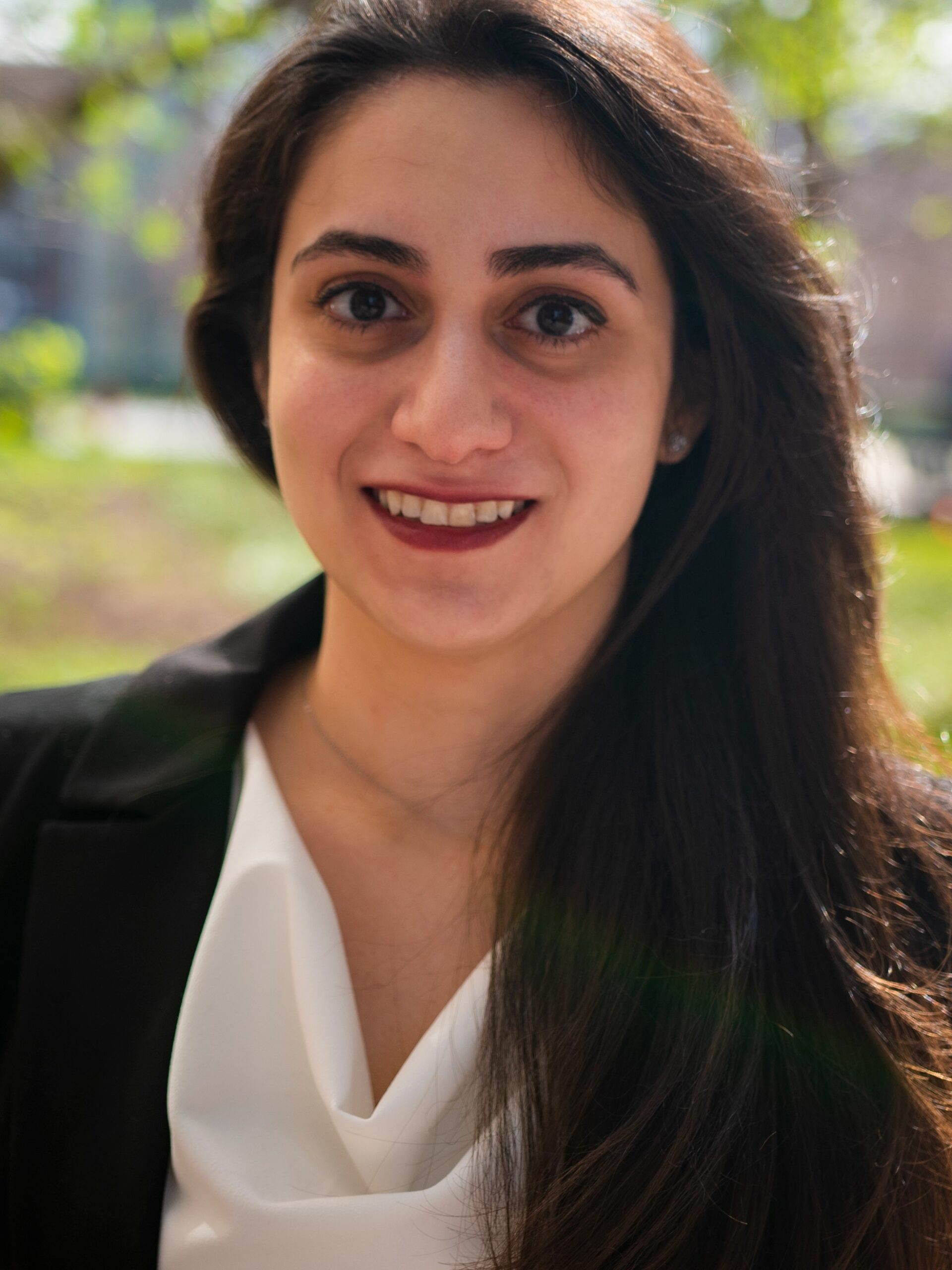
Former postdoctoral fellow and current professional research associate (medical imaging, University of Saskatchewan), Ghazal Azarfar, applied DL techniques to uterine and endometrial cancers with a similar goal of developing a fast and reliable IR technique for informing surgical resection of tumors with Bhargava and Georgina Cheng, M.D., from Carle Foundation Hospital.
“When a surgeon removes cancerous tissue from a patient with uterine cancer, they try to determine if there is any invasion into other organs. If there is, they must remove even more tissue. There is only 30-minutes for the on-call pathology team to assess the sample and help the surgeon,” Azarfar said. “With a robust IR computer-aided system, this task can be completed in the surgery room at a lower cost. Use of such systems may also help minimize the anesthesia time of the patient.”

Ghazal Azarfar.
Azarfar’s main challenge in this project was a lack of datasets for endometrial cancers. The issue persists for many cancer types with a lower mortality rate, especially with chemical imaging modalities, which remain a research technology.
However, the Bhargava lab’s students and researchers remain confident that with increasing collaborations with experts in other fields, including pathologists, clinicians, and cancer biology researchers, traditional lab procedures can be circumvented using AI methods.
“The discoveries and innovations that happen in my lab could not happen without the dedication and contributions of the incredible students and postdoctoral researchers. They bridge the gap between labs of different disciplines and departments, and they teach each other — and myself — new things every day. They are a driving force of much of the progress made in cancer research at the university,” said Bhargava.
– Written by the CCIL Communications Team
Rohit Bhargava is the Director of the Cancer Center at Illinois and Founder Professor of Engineering. His lab, the Chemical Imaging and Structures Laboratory, has pioneered the field of chemical imaging by developing optical theory for spectroscopy, new imaging instruments, AI tools, applications in pathology and methods for 3D tumor mimicking systems. Their work spans many cancers, including those of the breast, prostate, colon, brain, and skin.
* The collaboration with Anna Higham, M.D., is funded by the Cancer Center at Illinois’ Cancer Scholars for Translational and Applied Research (C★STAR) program. This funding was awarded to Anirudh Mittal, a graduate student in the Bhargava lab.

