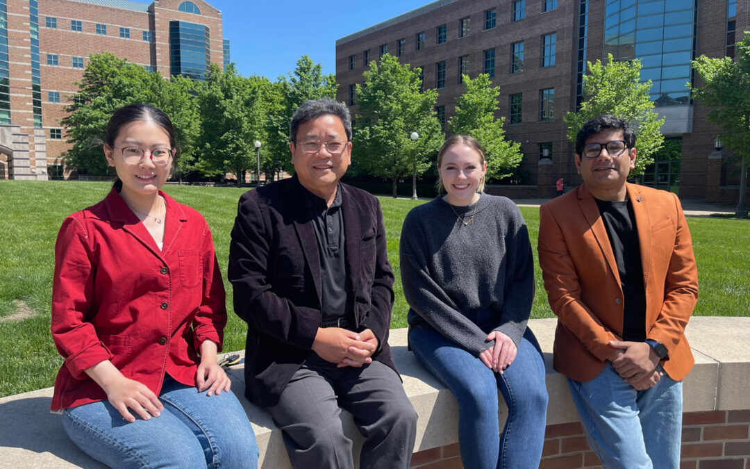
From left to right: Ruiyang Xue, Shuming Nie, Jamie Jones, and Indrajit Srivastava.
Urbana, Ill. – Surface-enhanced Raman scattering (SERS) gold nanoparticles are an ultrasensitive tool used by researchers for in-vitro medical diagnostics and for providing crucial information during cancer surgery. Cancer Center at Illinois (CCIL) researchers have developed a method to increase the storage and transport life of such nanoparticles to improve their utility in labs and clinics.
SERS nanoparticles are based on a class of plasmonic nanoparticles, which have free electrons that can be excited via a laser beam. This excitation amplifies the electromagnetic field at the surface of the nanoparticle and enables imaging technologies to pick up signatures, illuminating target biological features such as cancer cells.
However, current gold nanoparticles have limited use as they aggregate quickly and cannot be frozen or lyophilized (freeze dried) for later use or transport to remote locations. An Illinois team comprised of primary author and postdoctoral fellow Indrajit Srivastava and his co-supervisors Shuming Nie and Viktor Gruev has developed a solution using red blood cell membranes (RBCM) as a biomimetic surface coating.
The study, “Biomimetic Surface-Enhanced Raman Scattering Nanoparticles with Improved Dispersibility, Signal Brightness, and Tumor Targeting Functions,” was published in ACS Nano, a peer-reviewed journal of the American Chemical Society (ACS).
“I started working on plasmonic nanoparticles during my postdoc, but their instability has been an issue for quite some time. Several coating strategies have been developed thus far but have not been able to allow plasmonic nanoparticles to be frozen or lyophilized into powders, which would enable efficient transportation and storage. I explored different coatings that would improve the dispersibility of plasmonic nanoparticles but would also impart with important characteristics,” Srivastava said.
Inspired by nature, the team used a coating strategy by preparing membrane fragments of red blood cells using a bath sonication treatment. The fragments were then optimized to facilitate binding to the plasmonic surface of the nanoparticles of different shapes and sizes, forming a biomimetic coating.
“Indrajit had an idea to merge the nanoparticles and the natural cell membranes, and it worked very well in the lab, not only improving the stability and biocompatibility of the particles, but also increasing the brightness, or SERS signal intensity, when we imaged them,” said Nie, CCIL scientist and professor of bioengineering.
Standard SERS nanoparticles are stored in aqueous solutions, but the team’s biomimetic coating would allow researchers to prepare nanoparticles in powder form, enabling longer-term storage and shipping that could enable more collaborations between research institutions.
The team further demonstrated that such biomimetic nanoparticles could be easily modified by peptides to target cell-surface cancer biomarkers. They were able to verify their selective uptake in tumor cells via SERS spectroscopy and darkfield microscopy. The advances made with this development represent an important step towards clinical applications, especially for cancer diagnostics and in vivo imaging, including spectroscopy-guided cancer surgery.
The group believes a similar strategy could be used to develop biomimetic nanoparticles by using membranes from cancer and immune cells, opening new possibilities in developing nanoparticle vaccines and cancer immunotherapy.
– Written by the CCIL Communications Team
Shuming Nie is a Grainger Distinguished Chair and professor of Bioengineering. Viktor Gruev is a professor of electrical and computer engineering. Indrajit Srivastava is a postdoctoral research associate in the Nie lab and is co-supervised by Nie and Gruev. Other team members contributing to this study include Ruiyang Xue, graduate student in materials science and engineering; Jamie Jones, undergraduate student in bioengineering; Hyunjoon Rhee, undergraduate student in industrial engineering; and Kristen Flatt, staff scientist in the Materials Research Lab.-
The paper, “Biomimetic Surface-Enhanced Raman Scattering Nanoparticles with Improved Dispersibility, Signal Brightness, and Tumor Targeting Functions,” is available online. DOI: 10.1021/acsnano.2c01062.

