Urbana, Ill. — Cancer Center at Illinois (CCIL) member, Michael Oelze, and his collaborative breast cancer and ultrasound imaging research with Sunnybrook Health Sciences Centre in Toronto, Canada, was awarded a major $2M grant from the National Cancer Institute (NCI).
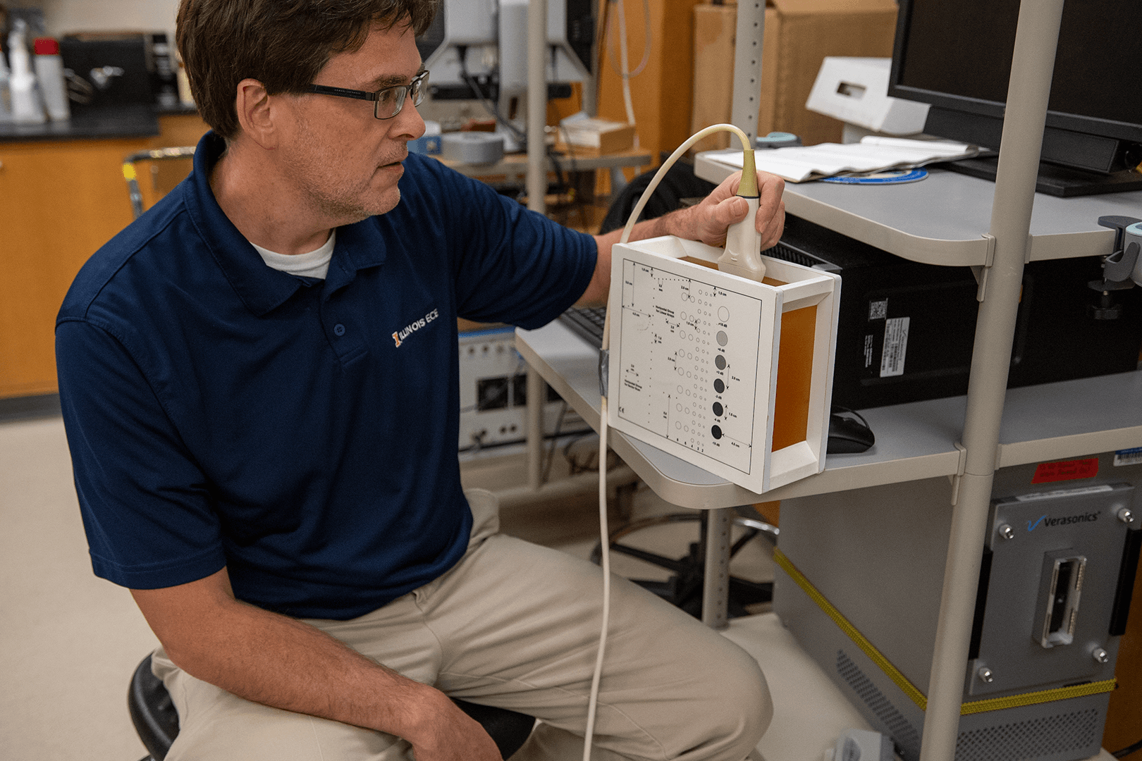
Oelze, Illinois professor of Electrical and Computer Engineering and Carle Illinois College of Medicine, is working with Professor Gregory Czarnota (MD, PhD) on a project that aims to identify the early response of breast cancer patients to neoadjuvant chemotherapy using quantitative ultrasound imaging technology.
Currently, it can take up to a few months to identify if a cancer treatment is working, but Oelze and collaborators hope to detect changes within a week. Radiological clips are inserted into tissue to help researchers locate, image, and determine if changes in the tumor are significant based on ultrasonic backscatter from the clips and surrounding tumor tissue.
Changes can occur in the signal from tumors due to a response to treatment or due to differences in how the image is taken from one scan session to the next. By utilizing radiological clips embedded in the tumors, differences in signals (related to how the image was taken) can be removed. This can result in better accuracy of quantitative ultrasound imaging to detect tissue signal changes.
“The current [project] isn’t looking at trying to diagnose breast cancer, it’s looking at how early and how well we can detect early response of breast cancer patients to neoadjuvant chemotherapy,” said Oelze.
The ultrasound detects whether the cancer cells are undergoing cell death by their shape and ultrasound backscattered signature. Changes in shape, size, and structure of cells within the tumor help researchers determine if the therapy is working.

Machine learning has played a role in this research by picking up signals that models used in previous studies had missed. In the past, image processing models have been used to analyze the image signals to assess masses and identify cell death. These models alone are helpful for interpreting the image, but the addition of machine learning allows for a more accurate assessment of the patient’s response by picking up on features that had been missed.
“We are planning on [a study of] around 150 patients. They’ll have multiple 3-dimensional scans of the tumors, multiple image frames we can look at, and we’ll scan them multiple times at baseline (i.e., just before the onset of chemotherapy), one-week post-therapy, two weeks, four weeks, and so forth,” said Oelze.
While the CCIL is home to many labs and research, clinical trials are not currently conducted on the U of I campus, making collaborations with local hospitals and organizations essential to research progress. Oelze credits the CCIL with helping him establish connections at a local hospital to test imaging techniques.
“The College of Medicine, the CCIL, and all these things combined will spur this sort of clinical research and collaboration that we really need here at Illinois,” said Oelze.
Oelze is currently working on other collaborative projects for treating tumors with ultrasound-based therapy techniques. His lab members are also working on detecting micro-calcifications in lesions and tumors to improve detection rates in cancer patients.
— Written by Olivia Fleming, Communications Intern

