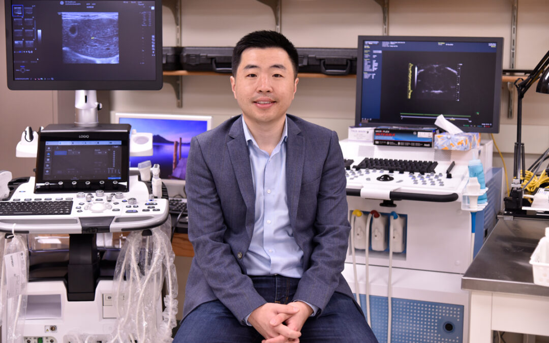
Urbana, Ill. – Pengfei Song, an assistant professor of electrical and computer engineering, has received a National Institute of Biomedical Imaging and Bioengineering Trailblazer R21 Award for pursuing the next-generation ultrasound localization microscopy. The award provides an opportunity for early-career investigators to explore highly innovative research projects.
Song is the principal investigator for the project, which was awarded more than $565,000. The research will build on technology that provides very high spatial resolution for imaging microvessels deep within tissue.
The imaging-based detection and characterization of abnormal tissue microvascular variations could be clinically significant for diagnosis, treatment evaluation, and therapy development in many pathologies such as cancer, cardiovascular diseases, inflammation, and neurodegenerative diseases. Currently, no viable noninvasive imaging tool can fulfill this important clinical need.
To fill this gap, Song’s project proposes to develop a new ultrasound-based super-resolution microvessel imaging technique that can directly and quantitatively assess the structure and the function of tissue microcirculation in vivo in humans.
“Ultrasound localization microscopy is a relatively young technology that many groups, including our group, have been actively working on,” Song said. “It stands out among other biomedical imaging modalities because it produces spectacular images of tissue microvasculature in vivo at depth. No other imaging modalities image as deep as ULM while offering the same level of spatial resolution as ULM. As such, ULM rapidly gained traction and found many biomedical applications such as cancer and neural imaging.
“However, as a young technology, ULM still has lots of technical barriers that we need to overcome to make it a truly useful technology for both basic science research and clinical application.”
Song said that ULM currently is an expensive (computationally) and slow technology.
“It is an ultrasound-based imaging method but it is not real-time, which is a major drawback for integrating ULM into the clinical ultrasound imaging workflow,” Song said. “In addition, ULM is mostly done in 2D at the moment, but we all know that anatomy and tissue blood flow are 3D. Having no access to 3D limits the accuracy of ULM imaging and makes it less useful for comprehensive evaluation of tissue microvasculature.”
The overall objective of the research in the proposal is to target these specific technical barriers of ULM, and to leverage the power of deep learning and new 3D ultrafast ultrasound techniques to overcome them.
– Written by the Beckman Institute

