Urbana, Ill. – Artificial intelligence (AI), algorithms that teach computers to perform tasks, can efficiently identify patterns and relationships in large amounts of data. These abilities have made AI techniques and tools indispensable in the cancer research community, especially for Cancer Center at Illinois (CCIL) scientists who are developing improved imaging tools used to diagnose cancers and guide decisions made in the clinical workflow.
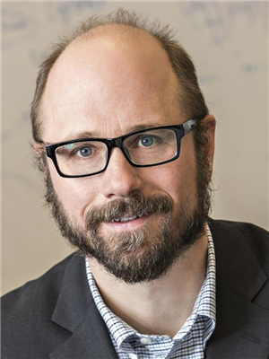
For Mark A. Anastasio, CCIL scientist and head of bioengineering, machine learning (ML) and deep learning (DL) methods are especially useful for solving a variety of imaging problems that affect many different imaging modalities and processes. His lab is readily adopting and developing new ML and DL methods to improve image reconstruction and analysis to better aid physicians and fellow scientists in making inferences about cancer and other diseases.
“When we talk about using AI to help us form or reconstruct better images, one of the first things we think about is how we can blend the physics of the imaging system and AI/ML,” Anastasio said. “We take what we know about the data and combine that with a suitable ML method to help us form images that will be useful for a specific diagnostic task.”

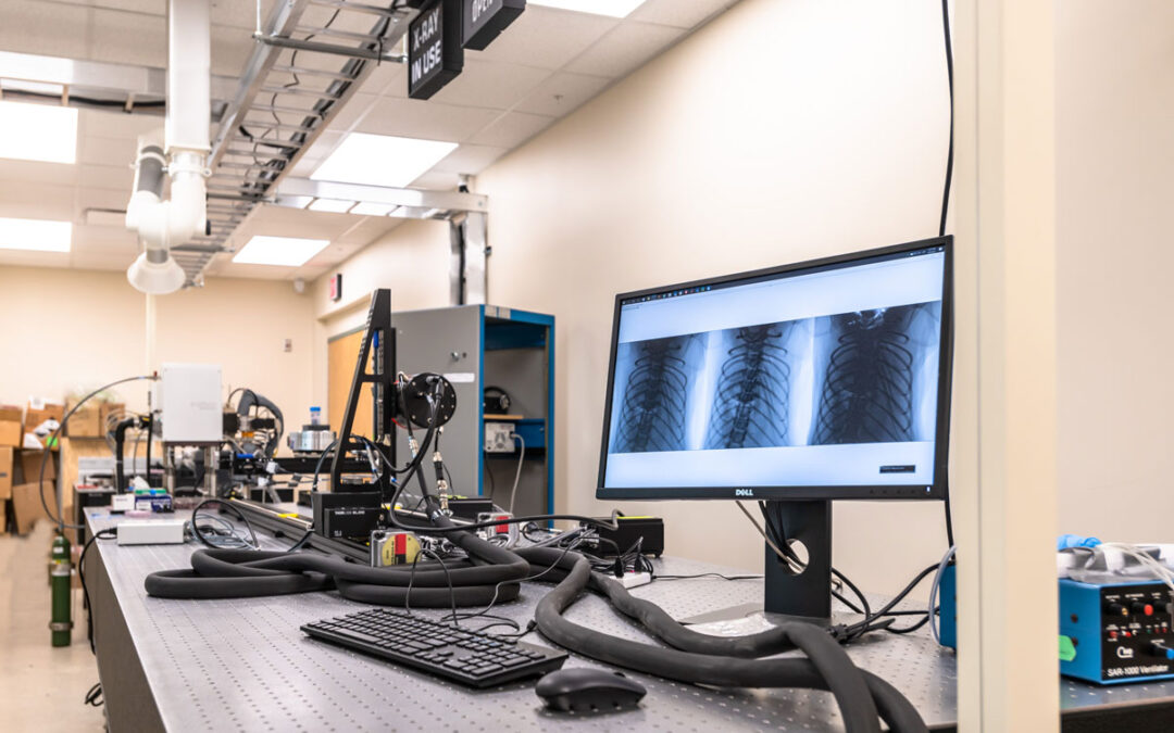
Imaging systems such as MRI and CT scanners acquire physics-based measurements about the patient that depend on biological features such as tissue characteristics; these measurements must then be reconstructed via computational algorithms that process the input data to form an image. AI methods are opening many new avenues for improving these processes, such as mitigating errors or compensating for incomplete or missing measurements.
The images obtained can be assessed by objective, task-based measures of image quality. For example, a medical image may be determined to be “good” if it is useful to a physician for tumor detection.
“At the end of the day, that’s what’s important. So, we’re also using DL to assess figures of merit that assess these types of image quality measures, and then we can better optimize diagnostic tasks, including cancer detection,” Anastasio said.
Anastasio’s lab uses neural networks, non-linear mappings between sets of inputs and outputs, to take their AI research even further. In particular, convolutional neural networks (CNN) are extremely well suited for use with image data, as these networks can learn how to discover features of data that are important for making decisions. Previously, these features had to be “hand-engineered,” or manually identified by the researcher, and fed to the AI.
These types of networks are commonly used in facial recognition software and are now ubiquitously used in the medical imaging field for image analysis and segmentation, predicting treatment outcomes, and cancer diagnosis, in addition to many other medical tasks.
Anastasio’s expertise with AI is instrumental in collaborative projects with other CCIL scientists Gabriel Popescu (professor of electrical and computer engineering), Stephen Boppart (CCIL program leader; professor of engineering), and Andrew Smith (associate professor of bioengineering). ML methods are being applied to improve their respective microscopy tools and techniques, in turn leading to better image segmentation and identification of biological structures such as cancer cells. The Anastasio lab is also currently working towards improving ultrasound tomography for better detection and diagnosis of breast cancer, funded by the NIH.
Widespread use of AI in the clinic is currently hindered by several issues. All new medical methods and tools must be regulated by the FDA, and as a new technology, ML methods require approval. Further, robust training of DL methods requires an incredible amount of data that must be curated to be representative of the entire population and contain the pathologies that could be encountered. This is especially important within a medical context, so all patients may benefit.
“I think we have room to grow in terms of the breadth of problems that we are addressing, but for imaging problems that are being addressed by AI and computational methods, the University of Illinois is definitely at the forefront of research,” Anastasio said. “More specifically, in terms of applying those methods to cancer, it is at the cutting edge.”
– Written by the CCIL Communications Team
Mark Anastasio is a member of the Cancer Center at Illinois, Donald Biggar Willet Professor in Engineering, and head of the department of bioengineering at the University of Illinois Urbana-Champaign.
Click here to read more about Anastasio’s lab, the Computational Imaging Science Laboratory.

