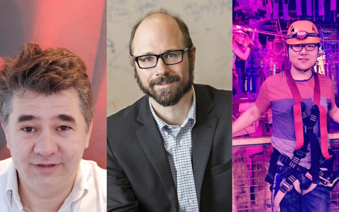
Left to right: Gabriel Popescu, Mark A. Anastasio, Chenfei Hu
Treating cancer patients and developing biopharmaceutical drugs depends on making the distinction between a healthy live-cell and a sickly dead cell. The state of the cell can indicate whether a cancer treatment is working or a new type of medicine is effective.
Groundbreaking research from the Beckman Institute for Advanced Science and Technology at the University of Illinois Urbana-Champaign can determine the state of a cell without the limitations inherent to some current methods.
“Current techniques for assessing cell viability involve dyes or stains that themselves kill the cells in a matter of minutes. It is almost like taking the pulse of a patient with a razor blade. Our approach now allows for a non-destructive assessment, which can be applied over arbitrary time scales, in real-time,” said Gabriel Popescu, a professor of electrical and computer engineering and a lead investigator on this study.
ECE alumnus Chenfei Hu, a Ph.D. candidate at the time of this research, led his team to develop an entirely new, label-free method of determining whether a cell is dead or alive. The study was published in Nature Communications in February 2022.
“Traditionally, people inject certain chemicals to determine the condition of the cells, which means a portion of the cells has to be sacrificed,” Hu said.
Using the current methods, researchers are unable to study cells at an individual level. Due to the sacrificial nature of chemically labeling a portion of the cells, it is difficult to study the entirety of the cells over a long period of time.
Hu’s research, which began in 2020, combines optimal imaging and machine learning to introduce the live-dead assay method, which uses advanced imaging technology and computational deep-learning algorithms to determine the state of the cell without chemical staining. This is made possible as the machine-learning technology studies images of cells on an individual level and learns how to spot a healthy versus an unhealthy cell.
“Using this combined technology, we can look at these cells continuously, over long periods of time, which cannot be done in the traditional way,” Hu said. “This is something nobody has done before, and also a way to save a lot of materials.”
Hu’s team has found such success with this new method, which achieved a 95% accuracy rate, that they have applied for a patent — the first step in bringing the method into real-world settings, which could include hospitals, additional research labs, and the broader medical community.
Like most research conducted within the Beckman Institute, this study was made possible through cross-disciplinary collaboration.
“This development has been [made] possible by combining expertise in label-free imaging from our lab with that in deep learning from Mark Anastasio’s group,” Popescu said.
Mark Anastasio is a professor of bioengineering and the head of the Department of Bioengineering at the University of Illinois.
“This research is an exciting example of how imaging physics, advanced instrumentation, and deep learning can be integrated to address impactful problems that were intractable only a few years ago,” Anastasio said.
The paper associated with this work can be accessed at https://doi.org/10.1038/s41467-022-28214-x
– Written by

