
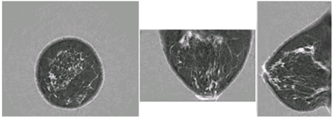
Images showing coronal, axial, and sagittal speed of sound images of the breast, taken with the QT Imaging scanner.
Urbana, Ill. – A Cancer Center at Illinois (CCIL) researcher has been awarded a multi-million-dollar federal grant to develop an advanced ultrasound-based imaging system for monitoring breast cancer patients’ response to chemotherapy. The work is a collaboration with industry and clinical researchers to produce higher-quality, timely, and detailed imaging data for breast cancer that ultimately contributes to improved patient outcomes.
Michael Oelze, CCIL scientist and Carle Illinois Health Innovation Professor, is leading the project, pairing a unique quantitative ultrasound technology he developed at the University of Illinois Urbana-Champaign with a table-mounted scanner developed by industry partner QT Imaging. The QT Ultrasound® is already FDA-approved to supplement mammography for diagnostic purposes, producing images of the breast with state-of-the-art contrast. A nearly $2.6 million, five-year grant from the National Institutes of Health and the National Cancer Institute allows Oelze and his team to combine efforts with QT Imaging and use machine learning to produce a unique breast-scanning system that will improve cancer monitoring.
“The integration of our technology onto the QT Imaging platform is the perfect marriage of our technologies,” said Oelze, who is a professor in the Department of Electrical and Computer Engineering at UIUC and is affiliated with the Beckman Institute for Advanced Science and Technology.
Most breast cancer patients undergo initial treatment with either chemotherapy or radiation to shrink the size of their tumors prior to surgery. But current monitoring methods – including traditional hand-held ultrasound — often fail to detect early progression or tumors that don’t respond to initial treatment. “Our preliminary data suggest that we should be able to identify response of patients to chemotherapy within a week of therapy onset,” Oelze said. “This early identification of response could provide clinicians with the information needed to adjust therapy if it is found that the current regimen is not working.”

Image of Michael Oelze.
The QT Imaging scanner produces images that are not only clearer, but are also quantitative, mapping the speed of the sound of ultrasonic waves passing through breast tissue. The data can be used to identify tissue characteristics and biomarkers that improve the precision of ultrasound findings. “We can improve the spatial resolution of our quantitative methods by four to five times by implementing our techniques on the QT Imaging scanner,” Oelze explained. “Their images also provide the ability to correct for attenuation and refraction artifacts (alterations in the image), a feature not available in traditional ultrasound.” The scanner is mounted in a special examination table to reduce alterations that can occur with movement and angle of hand-held ultrasound devices.
The new system will also be safe and cost-effective. No radiation or contrast dye is used in the test. “The final technology will be inexpensive and accurate, competing well with MRI/CT for breast cancer, driving down medical imaging costs by up to 90%,” Oelze said. Earlier data on progression or tumor response may also eliminate the need for additional biopsies and other interventions to determine if treatment is working.
Clinical partners at Sunnybrook Health Sciences Centre in Canada will test the effectiveness of the new device by scanning 100 to 120 breast cancer patients and then comparing their advanced quantitative ultrasound findings with the tumor response at the time of surgery.
Editor’s note: Michael Oelze is a Cancer Center at Illinois scientist, Carle Illinois Health Innovation Professor, and director of the college’s Capstone Program. He is also the Frederick G. and Elizabeth H. Nearing Scholar, professor and associate head for graduate affairs in the Department of Electrical and Computer Engineering and the Department of Bioengineering in The Grainger College of Engineering. He is an affiliate of Beckman Institute, Coordinated Science Laboratory, and Holonyak Micro & Nanotechnology Lab at the University of Illinois Urbana-Champaign.
Written by Beth Hart.